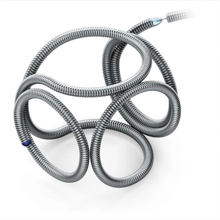
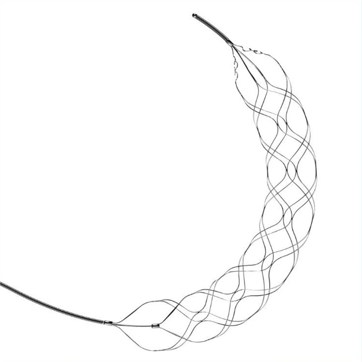
Subarachnoid hemorrhage(SAH) is a type of stroke, caused by bleeding in the space between the brain and the thin tissue that covers it. It is a serious and life-threatening condition that requires immediate medical attention. RenovaTM subarachnoid hemorrhage endovascular coiling is one of the treatment methods for SAH. In this method, a small metal coil is placed into the aneurysm(weakened blood vessel) that is causing the hemorrhage to stop the bleeding and prevent further damage.
Renova Subarachnoid Hemorrhage Endovascular Coiling is intended to endovascularlyobstruct or occlude blood flow invascular abnormalities of the neurovascular and peripheral vessels. It is indicated for endovascular mbolization of intracranial aneurysms, other neurovascular abnormalities such as arteriovenous malformations and arteriovenous fistulae, arterial and venous embolizations in the peripheral vasculature.
Features &Advantages
1. Open loop configuration conforms to differentaneurysm shapes and minimizes compartmentalization.
2. Detachment tactile sense and visual sense duplex feedback, more reliable.
3. Hybrid delivery shaft features a balanced flexibility and pushability, smooth delivery.
4. From frame to finish, various softness levels and sizes cover a variety of cases.
5. Frame securely, fill uniformly and finish by seeking voids within the aneurysm.
Firstly, push the microcatheter into the targeted aneurysm. Draw back the micro-guiding wire. At the proximal of coil system, 10 cm breakable zone configurated on the push shaft. The push shaft is a stiffness and gradient structure. Coil equipped on the distal end of push shaft, which make sure it could be smoothly delivered, and greatly minimizes kick-back microcatheter. Before use, please check the coil system. Firstly, check whether it is broken or not at the proximal zone. Take the coil out. Bring the coil out of protective sheath and check the coil and detachable zone.3D coil has good performance for frame, due to the softness.
If there is no problem. Secondly, withdraw the coil into sheath again if there is no broken. Insert the sheath into the Y-RHV, reaching the half length of Y-RHV, and rotate Y-RHV tightly. Flush the sheath with a high-pressure infusion set. Until the saline overflows from the proximal end of the sheath. Loosen the Y-RHV and continue to insert sheath into the hub of microcatheter, and then fix and rotate it tightly. Push the sheath into the microcatheter. Make sure the distal segments of protective sheath and microcatheter is close tightly. Otherwise, the coil will be possible to be damaged at the distal part of microcatheter. Push shaft smoothly, deliver the coil into microcatheter. Loosen the Y-RHV. And withdraw the sheath from Y-RHV. Then tighten the Y-RHV properly to prevent blood backflow. Continue to push shaft smoothly. Two markers located at the proximal end, 145cm, 155cm from the distal of push shaft separately, which indicate that the coil is about to reach the targeted aneurysm under the fluoroscopic guidance. If the coil does not have frame well. It can be withdrawn, and then released again. When the marker of guide wire and the proximal one of microcatheter are moving like a “T” shape under fluoroscopic guidance, the coil can be detached by breaking the breaking zone of shaft. Then, pull the wire gently, the movement of detaching marker is visual under fluoroscopic guidance. At the same time, it can be felt that the load force released suddenly. Withdraw the guide wire slowly. Coil detached and released successfully. Then, withdraw the wire from the microcatheter quickly. Continue to use next coil as above steps. Until the expected filling effect achieved.