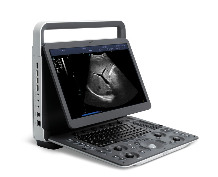
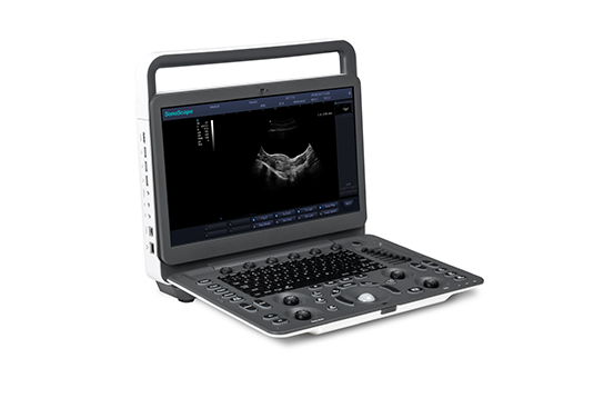
Cetus 40
Ultrasound Diagnostic System
-High-definition 21.5-inch monitor
-13.3-inch high sensitive touch screen
-Adjustable and intuitive control panel without keyboard
-Gel warmer (two temperature options)
-High-definition 21.5-inch monitor
-13.3-inch high sensitive touch screen
-Adjustable and intuitive control panel without keyboard
-Cable management solution
-Built-in battery for scanning
fAssist
fShare
fRemote
DICOM 3.0
Auto Optimization
Advanced Applications:
Panoramic
The extended field of view displays more image information without sacrificing image quality. A convenient approach for big-size organs, especially for MSK structures. It has scanning direction and speed indications.
Contrast Imaging
Pulse inversion contrast-enhanced ultrasound imaging technology can accurately extract the second harmonic of contrast microbubbles, realize contrast-enhanced imaging with high contrast-to-tissue ratio, and provide more detailed diagnosis for clinic.
Elastography
Real time elastography is a new noninvasive and painless technique that can help determine the hardness of organs and other structures such as breast, thyroid. Elastic imaging provides users with dynamic visual information and displays the rigidity of organs, which is helpful for direct and quantitative diagnosis and treatment.
AM
It permits the M-mode cursor to be angled in any direction on digital two-dimensional images. The three sample lines make the doctor measure three positions at the same time and each sample line can be rotated up to 360°
Curved AM
Curved Anatomical M-Mode (CAM) technology can show all the spatial and temporal relationship of myocardial segment movements during the cardiac cycle in the scanning sector, which provides a new measurement method to quantitatively analyze the abnormalities of segmental myocardial motion during systolic or diastolic period.
Tissue Doppler Imaging
Tissue Doppler Imaging (TDI) is a robust and reproducible echocardiographic tool that employs the Doppler effect to assess muscle wall characteristics throughout the cardiac cycle including velocity, displacement, deformation, and event timings. It has permitted a quantitative assessment of both global and regional function and timing of myocardial events.
Biopsy
Three guiding lines to ensure accuracy and safety. Width between two outer lines can be adjusted to fit the lesion areas, and needle depth information is available.
eBiopsy+
The image of the puncture needle is enhanced by the deflection of the acoustic beam, including needle enhancement, needle tip red rendering, virtual needle passage and scale line, supporting auto steering.
CW
Used to measure high-velocity blood flow inside the heart, obvious advantages for quantitative analysis of stenosis and regurgitation.
Smart Workflow:
Auto Volume Flow
Measure the blood vessel area, the blood flow velocity could be measured by spectrum automatically, then the blood flow volume results will show.
Auto IMT
Automatic identification and measurement of intima-media thickness. Both left and right blood vessels, anterior and posterior walls can be measured.
Auto Track-C
Quickly ROI re-locating on blood vessel by one button. Reduce doctor’s workload of frequently adjusting ROI position & angle due to different vessel location, improve efficiency
fShare
Support sending information via E-mail to computer, mobile phone, pad, etc. No software application is required on personal devices.
fAssist
Providing tutorial information regarding to abdomen, vascular, small parts, GYN, MSK, etc., including standard ultrasound image, anatomical diagram, scanning technique and tips.
Shortcut Key Tips
Shortcut key function displays in the bottom left corner,help doctors quickly locate required functions.
* Transducers :-
1) Convex C6-1s
Applications:Abdomen,
Obstetrics, Gynecology
2) Convex C5-1
Applications:Abdomen,
Obstetrics, Gynecology
3) Micro-convex MC10-3
Applications:Pediatrics,
Cardiology
4) Intracavitary EC9-4
Applications:Obstetrics,
Gynecology, Urology
5) Convex Volume V6-2
Applications:Abdomen,
Obstetrics, Gynecology
6) Intracavitary Volume EV10-3
Applications:Obstetrics,
Gynecology, Urology
7) Linear L12-4
Applications:Small parts,
Vascular, MSK
8) Linear L17-5
Applications:Small parts,
Vascular, MSK
9) HD Linear L13-3
Applications:Small parts,
Vascular, MSK, Breast
10) Linear L25-10
Applications:Small parts,
Vascular, MSK
11) Intracavitary EC10-3
Applications:Obstetrics,
Gynecology, Urology
12) Phased Array P5-2
Applications:Cardiology,
Abdomen, TCD
13) Phased Array P8-2
Applications:Abdomen,
Pediatric cardiology
14) Phased Array P10-3
Applications:Abdomen,
Neonatal cardiology
15) Phased Array P5-1s
Applications:Cardiology,
Abdomen, TCD